


2 HUMAN BOne & JOINT NANUNG DANAR DONO, S.Pt. MP. Worldwide Class – SMAN 1 Teladan Yogyakarta
4 Preliminary growing foetus Bloodstream vessels infant Growing processes kids Cakra epiphyse Spongious bone adult cartilage
5 Bone ( Latin: “os” ) is a kind of hard endoskeletal ligament present in human and lots of vertebrate creatures. Bones support body structures, safeguard internal organ, and (along with muscles) facilitate movement will also be associated with cell formation, calcium metabolic process, and mineral storage. The bones of the animal are, with each other, referred to as skeletal. Bone includes a different composition than cartilage, and both originated from mesoderm. Bone is really a living, growing tissue. It’s a porous mineralized structure, comprised of cells, vessels, crystals of calcium compounds (hydroxyapatite), the share which varies based on bone types and regions. HUMAN BONE PHYSIOLOGY
6 You will find five primary kinds of bone cells in navicular bone. 1.OSTEOGENIC cells react to traumas, for example fractures, by providing rise to bone-developing cells and bone-destroying cells. 2.OSTEOBLASTS (bone-developing cells) synthesize and secrete unmineralized ground substance and therefore are present in regions of high metabolic process inside the bone. 3.OSTEOCYTES are mature bone cells produced from osteoblasts which have renedered navicular bone around themselves. These cells maintain healthy navicular bone by secreting enzymes and manipulating the bone mineral content additionally they control the calcium release in the navicular bone towards the bloodstream. 4.OSTEOCLASTS are large cells that break lower navicular bone. They are important to bone growth, healing, and remodeling. 5.The final kind of cells are bone-lining cells.

These are manufactured from osteoblasts across the the surface of most bones within an adult. Bone-lining cells are believed to manage the movement of calcium and phosphate into and from the bone. BONE CELL

7 1. With respect to the Bone Member. a.Skull. cranial & facial bones b.Body. sternum, ribs, vertebral column c.Extremities. upper & lower extremities. 2. With respect to the Bone Shape. Regular Shape. Lengthy/Pipe bone. humerus, tibia, femur, ulna, metacarpals. Lengthy/Pipe bone. humerus, tibia, femur, ulna, metacarpals. Flat bone. ribs, cranial bones, bones of shoulder girdle. Flat bone. ribs, cranial bones, bones of shoulder girdle. Short bone. the wrists and ankles Short bone. the wrists and ankles Irregular Shape. the bones from the vertebrae along with a couple of within the skull (maxilla & mandible). BONE DIVISION Infant has greater than 300 bones. Adult has 206 ones
8 3. With respect to the Bone Material Composer. a.Bone. Made up of Osteoblast which produces Osteoblast & Osteocytes. You will find three kinds of cartilage. Hyaline. exist in the joints, in the the surface of a bone, trachea, larynx, etc. Hyaline. exist in the joints, in the the surface of a bone, trachea, larynx, etc. Fibrocartilage. made up of bovine collagen ” floating ” fibrous intervertebral disc, etc. Fibrocartilage. made up of bovine collagen ” floating ” fibrous intervertebral disc, etc. Elastic. made up of elastic ” floating ” fibrous in the ear & epiglottis.S Elastic. made up of elastic ” floating ” fibrous in the ear & epiglottis.S b.Cartilage.

Made up of Condrocytes which produce Condrin as matrix cells. Chondrocytes produce matrix known as chondroblasts. They have the effect of appositional growth.
9 HYALINE CARTILAGE contains cells known as chondrocytes baked into a distinctive matrix that provides the tissue both strength and versatility. The chondrocytes housed within their spaces known as “lacunae”. Some spaces contain several chondrocytes. Also note the outdoors from the cartilage tissue is ligament known as “perichondrium”. Layers of “chondroblasts” are based in the inner layers. They produce new chondrocytes because they secrete matrix around themselves. Layers of “chondroblasts” are based in the inner layers. They produce new chondrocytes because they secrete matrix around themselves. Cartilage can grow. 1.in the outdoors (appositional growth) and a pair of.from inside (interstitial growth). 2.from inside (interstitial growth).
10 ELASTIC CARTILAGE The cartilage within the EAR contains many elastic fibers and it is therefore known as “elastic cartilage”. Elastic cartilage could be distinguished through the stain for elastin which brings the dense bundles. FIBRO-CARTILAGE Fibrocartilage is distinguished by very scattered, infrequent chondrocytes and bovine collagen fibers running within the matrix. Fibrocartilage is distinguished by very scattered, infrequent chondrocytes and bovine collagen fibers running within the matrix. Fibrocartilage available at the intervertebral disk.
12 The study of joints is known as arthrology. Study regarding the motion of the body is known as kinesiology. Bones are extremely rigid to bend. Flexible connective tissues from joints and enable movement. Some pot, also known as articulation (link between bones) is an item of contact. ARTHROLOGY
13 Structurally joints are called following (what kinds of connective tissues from the joint). Structurally joints are called following (what kinds of connective tissues from the joint). ” floating ” fibrous joints. the bones are held together by ” floating ” fibrous ligament that’s wealthy in bovine collagen fibers. No synovial cavity. ” floating ” fibrous joints. the bones are held together by ” floating ” fibrous ligament that’s wealthy in bovine collagen fibers. No synovial cavity. Cartilaginous joints: the bones are held together by cartilage. No synovial cavity. Cartilaginous joints: the bones are held together by cartilage. No synovial cavity. Synovial joints: the bones developing the joint possess a synovial cavity and therefore are u . s . by dense irregular ligament. Synovial joints: the bones developing the joint possess a synovial cavity and therefore are u . s . by dense irregular ligament. JOINT CLASSIFICATION
14 Functionally joints are called following (what amount of movement have i got at this joint). Functionally joints are called following (what amount of movement have i got at this joint). SYNARTHROSIS: an immovable joint ( sendi mati ). AMPHIARTHROSIS: a rather movable joint ( sendi kaku ). DIARTHROSIS: a freely movable joint. All diarthroses are synovial joints ( sendi gerak ).
15 A. Suture – We already met these, found only within the skull, made from dense ” floating ” fibrous CT (irregular) that’s a remnant from the membranous fetal “skull”. Some sutures are temporary and ossify to get synostosis (such as the one that was once backward and forward halves from the mandible). Within the seniors, all sutures may ossify. B. Gomphosis – Teeth in sockets (alveoli). Here, the dense (” floating ” fibrous) CT store the tooth within the alveolar bone is frequently arranged, and it is more particularly referred to as periodontal ligament. C. Synchondrosis – Bones are became a member of by hyaline cartilage. Examples are epiphyseal plates (a brief joint), or even the joint between your first seven ribs and also the sternum (might also ossify as we grow older). D. Syntosis – Former joint which has now closed by bony fusion. Examples are metopic suture between fetal two frontal bones or even the epiphyseal line once an epiphyseal growth plate has closed. The lines of fusion may obliterate. SYNARTHROSIS – IMMOVABLE JOINT These could be structurally ” floating ” fibrous or cartilaginous
16 A. Syndesmosis – Bones are became a member of by an interosseous membrane or perhaps a ligament made from dense ” floating ” fibrous CT (irregular within the situation from the interosseous membrane or regular within the situation of the ligament), with increased connecting CT than suture, permitting a little bit of movement. Example is interosseous membrane between tibia and fibula or radius and ulna, or interosseous ligament between distal tibia and fibula. B. Symphysis – Bones are held together with a fibrocartilaginous pad or disc. Examples are intervertebral joints (between physiques of vertebrae, the joints between articular facets are synovial – and they are not symphyses) and genital symphysis. AMPHIARTHROSIS – SLIGHTLY MOVABLE JOINT These could be structurally ” floating ” fibrous or cartilaginous
17 A. Gliding (straight line) movements are easy, the opposing surfaces slide past each other, as between carpals, between tarsals and between clavicle and sternum. B. Angular movements tend to be more complicated know these and exercise them: DIARTHROSIS – FREELY MOVABLE JOINT All diarthroses are structurally synovial, simply because they require a joint cavity to become freely movable. They are common through the body, and therefore are probably the most familiar kind of joint, present in many shapes. Flexion. Flexion. Extension. Extension. Hyperextension Hyperextension Abduction Abduction Adduction Adduction Circumduction. Circumduction.
18 DIARTHROSIS – FREELY MOVABLE JOINT All diarthroses are structurally synovial, simply because they require a joint cavity to become freely movable. They are common through the body, and therefore are probably the most familiar kind of joint, present in many shapes. Flexion – generally (although not always) lessens the position between articulating bones or structures, as with bending the elbow or putting the face towards the chest. Flexion – generally (although not always) lessens the position between articulating bones or structures, as with bending the elbow or putting the face towards the chest. Extension – generally (although not always) boosts the position between articulating bones, and brings it well to physiological position. Extension – generally (although not always) boosts the position between articulating bones, and brings it well to physiological position. Hyperextension – extension continues beyond physiological position, for example in tilting the mind back using the face toward the ceiling. Hyperextension – extension continues beyond physiological position, for example in tilting the mind back using the face toward the ceiling. Abduction – movement of the bone from the midline from the body Abduction – movement of the bone from the midline from the body Adduction – movement of the bone toward the midline from the body, brings it to physiological position. Adduction – movement of the bone toward the midline from the body, brings it to physiological position. Circumduction – a circular motion that mixes flexion, extension, abduction, adduction in succession. Circumduction – a circular motion that mixes flexion, extension, abduction, adduction in succession.
19 C. Rotational movements – bone moves in one plane around its axis. This is often right and left, as with turning the mind back and forth (indicating “no”). It is also medial or lateral with regards to a corner, as with the low or upper limb. Turning your feet “out” is lateral rotation in the hip, while turning your toes “in” is medial rotation in the hip. Pronation and supination are rotational movements restricted to the elbow where radius articulates with capitulum from the humerus. D. Special Movements at Diarthroses – Extra movements are possible additionally towards the fundamental ones formerly pointed out. These special movements occur at specific joints. See text for illustrations. Elevation versus. depression (like shoulder shrugging or mandible opening and closing), protraction versus. retraction (for example with shoulders or mandible), inversion versus. eversion (ankle movement, turns sole of feet inward and outward, correspondingly), dorsiflexion versus. plantar flexion (ankle movement, getting toes up toward the low leg versus. pointing foot lower, correspondingly), opposition (for example between your thumb along with other fingers), and lateral flexion for example using the trunk. DIARTHROSIS – FREELY MOVABLE JOINT
20 Sutures. Just because a suture is immovable, it’s functionally considered a synarthrosis. Some sutures are substituted with bone within the adult. This type of suture is known as synostosis. We already met these, found only within the skull, made from dense ” floating ” fibrous CT (irregular) that’s a remnant from the membranous fetal “skull”. Within the seniors, all sutures may ossify. Sutures. Just because a suture is immovable, it’s functionally considered a synarthrosis. Some sutures are substituted with bone within the adult. This type of suture is known as synostosis. We already met these, found only within the skull, made from dense ” floating ” fibrous CT (irregular) that’s a remnant from the membranous fetal “skull”. Within the seniors, all sutures may ossify. Syndesmoses. Example tibia/fibula. Since it permits slight movement, a syndesmosis is classed functionally being an amphiarthrosis. Syndesmoses. Example tibia/fibula. Since it permits slight movement, a syndesmosis is classed functionally being an amphiarthrosis. Gomphoses. The only real example would be the articulations from the roots from the teeth using the sockets from the alveolar processes from the maxillae and mandible. The dense ” floating ” fibrous ligament is known as the periodonatal ligament. This really is functionally considered a synarthrosis. Gomphoses. The only real example would be the articulations from the roots from the teeth using the sockets from the alveolar processes from the maxillae and mandible. The dense ” floating ” fibrous ligament is known as the periodonatal ligament. This really is functionally considered a synarthrosis. AMPHIARTHROSIS – SLIGHTLY MOVABLE JOINT They are joints that lack a synovial cavity & permit little if any movement
21 1. Ball-and-Socket Joints ( sendi peluru ) 2. Hinge Joints ( sendi engsel ) 3. Pivot Joints ( sendi putar ) 4. Saddle Joints ( sendi pelana ) 5. Planar joints ( sendi luncur/geser ) 6. Condyloid Joints or Ellipsoidal ( sendi kondiloid ) SUBTYPES OF DIARTHROSES According to structural classifications associated with the kinds of movements they permit
22 1. BALL-AND-SOCKET Joints- ( sendi peluru ) for example the ball-like the surface of one bone fitting right into a cuplike depression of some other bone. Egs. Shoulder and hip joints. Multiaxial. 2. HINGE Joints – ( sendi engsel ) the convex the surface of one suits the concave the surface of another. Eg. Knee, elbow, ankle, interphalangeal. Monaxial (uniaxial means one direction movement). 3. PIVOT Joints – ( sendi putar ) here the rounded or pointed the surface of one bone articulates having a ring created partially by another bone & partially with a ligament. This really is monaxial. Examples atlanto-axial joint, radioulnar joint:turns palm anteriorly and posteriorly.
23 4. SADDLE Joints – ( sendi pelana ) here the articular the surface of one bone is saddle-formed and also the articular top of the other suits the “saddle”. Eg. Carpometacarpal joint. Biaxial. 5. PLANAR joints – ( sendi luncur/geser ) the articulating surfaces are flat or slightly curved. Example are intercarpal joints, intertarsal joints, sternoclavicular joints, acromioclavicular joints, sternocostal joints, vertebrocostal joints. 6. CONDYLOID Joints – ( sendi kondiloid ) also known as ellipsoidal joint. The convex oblong-formed projection of 1 suits the oblong-formed depression of some other. Eg. Wrist and metacarpophalangeal joints. Biaxial.
24 1. Ball-and-Socket Joints ( sendi peluru ) 2. Hinge Joints ( sendi engsel ) 3. Pivot Joints ( sendi putar ) 4. Saddle Joints ( sendi pelana ) 5. Planar joints ( sendi luncur/geser ) 6. Condyloid Joints or Ellipsoidal ( sendi kondiloid ) Kinds Of SYNOVIAL JOINTS Based on direction from the movements.
25 1. Bone & Joint disorders. fracture, fissure, dislocation (luxations) 2. Infection 3. Necroses. Why? 4. Mineral Deficiency (Particularly Ca-P). Egs. milk fever 5. Impaired growth. X/O formed 6. Brittle bones. Lack of vit. D & mineral Ca-P 7. Osteo arthritis is localized “put on-and-tear” joint disease occurring normally in senior years, or might be secondary to injuries. 8. Rheumatic joint disease is really a systemic disease where your defense mechanisms attacks and destroys your joints (an autoimmune disorder). 9. Ankylosing spondilitis (my dad’s disease) causes fusing of axial elements. 10. Bone Cancer. milloma multiple, osteosarkoma 11. Joint discomfort 12. Homeostatic Imbalances – Understand that “joint disease” is an extremely generic term for any group of (possibly hundreds of) related joint and muscle disorders. 13. Etc. SKELETAL DISORDERS
26 “Arth-…” = joint “…-algia” = discomfort “…-ectomy” = elimination of “…-itis” = inflammation of So what is your opinion “synovitis” means? What about someone getting a “bursectomy”? Might your granny have “arthralgia”? MEDICAL TERMINOLOGY
2 HUMAN BOne & JOINT NANUNG DANAR DONO, S.Pt. MP. Worldwide Class – SMAN 1 Teladan Yogyakarta
4 Preliminary growing foetus Bloodstream vessels infant Growing processes kids Cakra epiphyse Spongious bone adult cartilage
5 Bone ( Latin: “os” ) is a kind of hard endoskeletal ligament present in human and lots of vertebrate creatures. Bones support body structures, safeguard internal organ, and (along with muscles) facilitate movement will also be associated with cell formation, calcium metabolic process, and mineral storage. The bones of the animal are, with each other, referred to as skeletal. Bone includes a different composition than cartilage, and both originated from mesoderm. Bone is really a living, growing tissue. It’s a porous mineralized structure, comprised of cells, vessels, crystals of calcium compounds (hydroxyapatite), the share which varies based on bone types and regions. HUMAN BONE PHYSIOLOGY
6 You will find five primary kinds of bone cells in navicular bone. 1.OSTEOGENIC cells react to traumas, for example fractures, by providing rise to bone-developing cells and bone-destroying cells. 2.OSTEOBLASTS (bone-developing cells) synthesize and secrete unmineralized ground substance and therefore are present in regions of high metabolic process inside the bone. 3.OSTEOCYTES are mature bone cells produced from osteoblasts which have renedered navicular bone around themselves. These cells maintain healthy navicular bone by secreting enzymes and manipulating the bone mineral content additionally they control the calcium release in the navicular bone towards the bloodstream. 4.OSTEOCLASTS are large cells that break lower navicular bone. They are important to bone growth, healing, and remodeling. 5.The final kind of cells are bone-lining cells. These are manufactured from osteoblasts across the the surface of most bones within an adult. Bone-lining cells are believed to manage the movement of calcium and phosphate into and from the bone. BONE CELL
7 1. With respect to the Bone Member. a.Skull. cranial & facial bones b.Body. sternum, ribs, vertebral column c.Extremities. upper & lower extremities. 2. With respect to the Bone Shape. Regular Shape. Lengthy/Pipe bone. humerus, tibia, femur, ulna, metacarpals. Lengthy/Pipe bone. humerus, tibia, femur, ulna, metacarpals. Flat bone. ribs, cranial bones, bones of shoulder girdle. Flat bone. ribs, cranial bones, bones of shoulder girdle. Short bone. the wrists and ankles Short bone. the wrists and ankles Irregular Shape. the bones from the vertebrae along with a couple of within the skull (maxilla & mandible). BONE DIVISION Infant has greater than 300 bones. Adult has 206 ones
8 3. With respect to the Bone Material Composer. a.Bone. Made up of Osteoblast which produces Osteoblast & Osteocytes. You will find three kinds of cartilage. Hyaline. exist in the joints, in the the surface of a bone, trachea, larynx, etc. Hyaline. exist in the joints, in the the surface of a bone, trachea, larynx, etc. Fibrocartilage. made up of bovine collagen ” floating ” fibrous intervertebral disc, etc. Fibrocartilage. made up of bovine collagen ” floating ” fibrous intervertebral disc, etc. Elastic. made up of elastic ” floating ” fibrous in the ear & epiglottis.S Elastic. made up of elastic ” floating ” fibrous in the ear & epiglottis.S b.Cartilage. Made up of Condrocytes which produce Condrin as matrix cells. Chondrocytes produce matrix known as chondroblasts. They have the effect of appositional growth.
9 HYALINE CARTILAGE contains cells known as chondrocytes baked into a distinctive matrix that provides the tissue both strength and versatility. The chondrocytes housed within their spaces known as “lacunae”. Some spaces contain several chondrocytes. Also note the outdoors from the cartilage tissue is ligament known as “perichondrium”. Layers of “chondroblasts” are based in the inner layers. They produce new chondrocytes because they secrete matrix around themselves. Layers of “chondroblasts” are based in the inner layers. They produce new chondrocytes because they secrete matrix around themselves. Cartilage can grow. 1.in the outdoors (appositional growth) and a pair of.from inside (interstitial growth). 2.from inside (interstitial growth).
10 ELASTIC CARTILAGE The cartilage within the EAR contains many elastic fibers and it is therefore known as “elastic cartilage”. Elastic cartilage could be distinguished through the stain for elastin which brings the dense bundles. FIBRO-CARTILAGE Fibrocartilage is distinguished by very scattered, infrequent chondrocytes and bovine collagen fibers running within the matrix. Fibrocartilage is distinguished by very scattered, infrequent chondrocytes and bovine collagen fibers running within the matrix. Fibrocartilage available at the intervertebral disk.
12 The study of joints is known as arthrology. Study regarding the motion of the body is known as kinesiology. Bones are extremely rigid to bend. Flexible connective tissues from joints and enable movement. Some pot, also known as articulation (link between bones) is an item of contact. ARTHROLOGY
13 Structurally joints are called following (what kinds of connective tissues from the joint). Structurally joints are called following (what kinds of connective tissues from the joint). ” floating ” fibrous joints. the bones are held together by ” floating ” fibrous ligament that’s wealthy in bovine collagen fibers. No synovial cavity. ” floating ” fibrous joints. the bones are held together by ” floating ” fibrous ligament that’s wealthy in bovine collagen fibers. No synovial cavity. Cartilaginous joints: the bones are held together by cartilage. No synovial cavity. Cartilaginous joints: the bones are held together by cartilage. No synovial cavity. Synovial joints: the bones developing the joint possess a synovial cavity and therefore are u . s . by dense irregular ligament. Synovial joints: the bones developing the joint possess a synovial cavity and therefore are u . s . by dense irregular ligament. JOINT CLASSIFICATION
14 Functionally joints are called following (what amount of movement have i got at this joint). Functionally joints are called following (what amount of movement have i got at this joint). SYNARTHROSIS: an immovable joint ( sendi mati ). AMPHIARTHROSIS: a rather movable joint ( sendi kaku ). DIARTHROSIS: a freely movable joint. All diarthroses are synovial joints ( sendi gerak ).
15 A. Suture – We already met these, found only within the skull, made from dense ” floating ” fibrous CT (irregular) that’s a remnant from the membranous fetal “skull”. Some sutures are temporary and ossify to get synostosis (such as the one that was once backward and forward halves from the mandible). Within the seniors, all sutures may ossify. B. Gomphosis – Teeth in sockets (alveoli). Here, the dense (” floating ” fibrous) CT store the tooth within the alveolar bone is frequently arranged, and it is more particularly referred to as periodontal ligament. C. Synchondrosis – Bones are became a member of by hyaline cartilage. Examples are epiphyseal plates (a brief joint), or even the joint between your first seven ribs and also the sternum (might also ossify as we grow older). D. Syntosis – Former joint which has now closed by bony fusion. Examples are metopic suture between fetal two frontal bones or even the epiphyseal line once an epiphyseal growth plate has closed. The lines of fusion may obliterate. SYNARTHROSIS – IMMOVABLE JOINT These could be structurally ” floating ” fibrous or cartilaginous
16 A. Syndesmosis – Bones are became a member of by an interosseous membrane or perhaps a ligament made from dense ” floating ” fibrous CT (irregular within the situation from the interosseous membrane or regular within the situation of the ligament), with increased connecting CT than suture, permitting a little bit of movement. Example is interosseous membrane between tibia and fibula or radius and ulna, or interosseous ligament between distal tibia and fibula. B. Symphysis – Bones are held together with a fibrocartilaginous pad or disc. Examples are intervertebral joints (between physiques of vertebrae, the joints between articular facets are synovial – and they are not symphyses) and genital symphysis. AMPHIARTHROSIS – SLIGHTLY MOVABLE JOINT These could be structurally ” floating ” fibrous or cartilaginous
17 A. Gliding (straight line) movements are easy, the opposing surfaces slide past each other, as between carpals, between tarsals and between clavicle and sternum. B. Angular movements tend to be more complicated know these and exercise them: DIARTHROSIS – FREELY MOVABLE JOINT All diarthroses are structurally synovial, simply because they require a joint cavity to become freely movable. They are common through the body, and therefore are probably the most familiar kind of joint, present in many shapes. Flexion. Flexion. Extension. Extension. Hyperextension Hyperextension Abduction Abduction Adduction Adduction Circumduction. Circumduction.
18 DIARTHROSIS – FREELY MOVABLE JOINT All diarthroses are structurally synovial, simply because they require a joint cavity to become freely movable. They are common through the body, and therefore are probably the most familiar kind of joint, present in many shapes. Flexion – generally (although not always) lessens the position between articulating bones or structures, as with bending the elbow or putting the face towards the chest. Flexion – generally (although not always) lessens the position between articulating bones or structures, as with bending the elbow or putting the face towards the chest. Extension – generally (although not always) boosts the position between articulating bones, and brings it well to physiological position. Extension – generally (although not always) boosts the position between articulating bones, and brings it well to physiological position. Hyperextension – extension continues beyond physiological position, for example in tilting the mind back using the face toward the ceiling. Hyperextension – extension continues beyond physiological position, for example in tilting the mind back using the face toward the ceiling. Abduction – movement of the bone from the midline from the body Abduction – movement of the bone from the midline from the body Adduction – movement of the bone toward the midline from the body, brings it to physiological position. Adduction – movement of the bone toward the midline from the body, brings it to physiological position. Circumduction – a circular motion that mixes flexion, extension, abduction, adduction in succession. Circumduction – a circular motion that mixes flexion, extension, abduction, adduction in succession.
19 C. Rotational movements – bone moves in one plane around its axis. This is often right and left, as with turning the mind back and forth (indicating “no”). It is also medial or lateral with regards to a corner, as with the low or upper limb. Turning your feet “out” is lateral rotation in the hip, while turning your toes “in” is medial rotation in the hip. Pronation and supination are rotational movements restricted to the elbow where radius articulates with capitulum from the humerus. D. Special Movements at Diarthroses – Extra movements are possible additionally towards the fundamental ones formerly pointed out. These special movements occur at specific joints. See text for illustrations. Elevation versus. depression (like shoulder shrugging or mandible opening and closing), protraction versus. retraction (for example with shoulders or mandible), inversion versus. eversion (ankle movement, turns sole of feet inward and outward, correspondingly), dorsiflexion versus. plantar flexion (ankle movement, getting toes up toward the low leg versus. pointing foot lower, correspondingly), opposition (for example between your thumb along with other fingers), and lateral flexion for example using the trunk. DIARTHROSIS – FREELY MOVABLE JOINT
20 Sutures. Just because a suture is immovable, it’s functionally considered a synarthrosis. Some sutures are substituted with bone within the adult. This type of suture is known as synostosis. We already met these, found only within the skull, made from dense ” floating ” fibrous CT (irregular) that’s a remnant from the membranous fetal “skull”. Within the seniors, all sutures may ossify. Sutures. Just because a suture is immovable, it’s functionally considered a synarthrosis. Some sutures are substituted with bone within the adult. This type of suture is known as synostosis. We already met these, found only within the skull, made from dense ” floating ” fibrous CT (irregular) that’s a remnant from the membranous fetal “skull”. Within the seniors, all sutures may ossify. Syndesmoses. Example tibia/fibula. Since it permits slight movement, a syndesmosis is classed functionally being an amphiarthrosis. Syndesmoses. Example tibia/fibula. Since it permits slight movement, a syndesmosis is classed functionally being an amphiarthrosis. Gomphoses. The only real example would be the articulations from the roots from the teeth using the sockets from the alveolar processes from the maxillae and mandible. The dense ” floating ” fibrous ligament is known as the periodonatal ligament. This really is functionally considered a synarthrosis. Gomphoses. The only real example would be the articulations from the roots from the teeth using the sockets from the alveolar processes from the maxillae and mandible. The dense ” floating ” fibrous ligament is known as the periodonatal ligament. This really is functionally considered a synarthrosis. AMPHIARTHROSIS – SLIGHTLY MOVABLE JOINT They are joints that lack a synovial cavity & permit little if any movement
21 1. Ball-and-Socket Joints ( sendi peluru ) 2. Hinge Joints ( sendi engsel ) 3. Pivot Joints ( sendi putar ) 4. Saddle Joints ( sendi pelana ) 5. Planar joints ( sendi luncur/geser ) 6. Condyloid Joints or Ellipsoidal ( sendi kondiloid ) SUBTYPES OF DIARTHROSES According to structural classifications associated with the kinds of movements they permit
22 1. BALL-AND-SOCKET Joints- ( sendi peluru ) for example the ball-like the surface of one bone fitting right into a cuplike depression of some other bone. Egs. Shoulder and hip joints. Multiaxial. 2. HINGE Joints – ( sendi engsel ) the convex the surface of one suits the concave the surface of another. Eg. Knee, elbow, ankle, interphalangeal. Monaxial (uniaxial means one direction movement). 3. PIVOT Joints – ( sendi putar ) here the rounded or pointed the surface of one bone articulates having a ring created partially by another bone & partially with a ligament. This really is monaxial. Examples atlanto-axial joint, radioulnar joint:turns palm anteriorly and posteriorly.
23 4. SADDLE Joints – ( sendi pelana ) here the articular the surface of one bone is saddle-formed and also the articular top of the other suits the “saddle”. Eg. Carpometacarpal joint. Biaxial. 5. PLANAR joints – ( sendi luncur/geser ) the articulating surfaces are flat or slightly curved. Example are intercarpal joints, intertarsal joints, sternoclavicular joints, acromioclavicular joints, sternocostal joints, vertebrocostal joints. 6. CONDYLOID Joints – ( sendi kondiloid ) also known as ellipsoidal joint. The convex oblong-formed projection of 1 suits the oblong-formed depression of some other. Eg. Wrist and metacarpophalangeal joints. Biaxial.
24 1. Ball-and-Socket Joints ( sendi peluru ) 2. Hinge Joints ( sendi engsel ) 3. Pivot Joints ( sendi putar ) 4. Saddle Joints ( sendi pelana ) 5. Planar joints ( sendi luncur/geser ) 6. Condyloid Joints or Ellipsoidal ( sendi kondiloid ) Kinds Of SYNOVIAL JOINTS Based on direction from the movements.
25 1. Bone & Joint disorders. fracture, fissure, dislocation (luxations) 2. Infection 3. Necroses. Why? 4. Mineral Deficiency (Particularly Ca-P). Egs. milk fever 5. Impaired growth. X/O formed 6. Brittle bones. Lack of vit. D & mineral Ca-P 7. Osteo arthritis is localized “put on-and-tear” joint disease occurring normally in senior years, or might be secondary to injuries. 8. Rheumatic joint disease is really a systemic disease where your defense mechanisms attacks and destroys your joints (an autoimmune disorder). 9. Ankylosing spondilitis (my dad’s disease) causes fusing of axial elements. 10. Bone Cancer. milloma multiple, osteosarkoma 11. Joint discomfort 12. Homeostatic Imbalances – Understand that “joint disease” is an extremely generic term for any group of (possibly hundreds of) related joint and muscle disorders. 13. Etc. SKELETAL DISORDERS
26 “Arth-…” = joint “…-algia” = discomfort “…-ectomy” = elimination of “…-itis” = inflammation of So what is your opinion “synovitis” means? What about someone getting a “bursectomy”? Might your granny have “arthralgia”? MEDICAL TERMINOLOGY

 Korapsyon sa pilipinas thesis writing
 Determinants of economic growth thesis writing
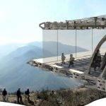 Digital architecture thesis proposal titles
 Ocad environment design thesis proposal
 Nspires change proposal title of thesis


