Special Issue: Spotlight on Cardiovascular Imaging
- R.J. Everett a,.
- C.G. Stirrat a
- S.I.R. Semple a,b
- D.E. Newby a,b
- M.R. Dweck a
- S. Mirsadraee a,b
- an english Heart Foundation/College Center for Cardiovascular Science, College of Edinburgh, United kingdom
- b Clinical Research Imaging Center, College of Edinburgh, United kingdom
Received 10 November 2015. Revised 15 The month of january 2016. Recognized 9 Feb 2016. Available on the web 19 March 2016.
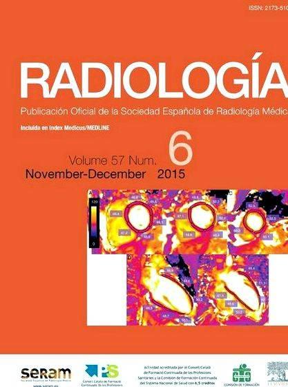
Myocardial fibrosis can arise from a variety of pathological processes and it is presence correlates with adverse clinical outcomes. Cardiac magnetic resonance (CMR) can offer a non-invasive assessment of cardiac structure, function, and tissue characteristics, including late gadolinium enhancement (LGE) strategies to identify focal irreversible substitute fibrosis having a high amount of precision and reproducibility. Importantly the existence of LGE is actually connected with adverse outcomes in a variety of common cardiac conditions however, LGE techniques are qualitative and not able to identify diffuse myocardial fibrosis, that is an early on type of fibrosis preceding substitute fibrosis which may be reversible. Novel T1 mapping techniques allow quantitative CMR assessment of diffuse myocardial fibrosis with two of the most common measures being native T1 and extracellular volume (ECV) fraction. Native T1 differentiates normal from infarcted myocardium, is abnormal in hypertrophic cardiomyopathy, and could be particularly helpful in detecting Anderson–Fabry disease and amyloidosis. ECV is really a surrogate way of measuring the extracellular space and is the same as the myocardial amount of distribution from the gadolinium-based contrast medium.
It’s reproducible and correlates well with fibrosis on histology. ECV is abnormal in patients with cardiac failure and aortic stenosis, and it is connected with functional impairment during these groups. T1 mapping techniques promise to permit earlier recognition of disease, monitor disease progression, and inform prognosis however, limitations remain. Particularly, reference ranges are missing for T1 mapping values because these suffer from specific CMR techniques and magnetic field strength. Additionally, there’s significant overlap between T1 mapping values in healthy controls and many disease states, particularly using native T1, restricting the clinical use of they at the moment.
Figure 1. Figure 2. Figure 3.
Table 1. Figure 4. Figure 5.
Guarantor and correspondent: R. J. Everett, British Heart Foundation/College Center for Cardiovascular Science, Room SU 305 Chancellor’s Building, College of Edinburgh, 49 Little France Crescent, Edinburgh EH16 4SB, United kingdom. Tel. +44 0131 242 6515 fax: +44 0131 242 6379.
2016 The Royal College of Radiologists. Printed by Elsevier Limited. All legal rights reserved.
Citing articles ( )




 Unique ways of writing your name
 Black cat crosses your path right to left writing
 Writing a research question and hypothesis development
 Mysterium board game english rules for writing
 My lost city luc sante summary writing
