Our Guarantees Our Quality Standards Our Fair Use Policy
How Come United kingdom Essays Different?
- There is a verifiable exchanging history as being a United kingdom registered company (details within the finish of every page).
- Our Nottingham offices are suitable for purchase to everybody to satisfy we greater than 40 full-time staff.
- United kingdom Essays partner with Feefo.com to produce verified customer testimonials – both positive and negative!
Ask an expert FREE
Ask an expert Index Ask an issue Compensated Services
About Our Ask an expert Service
Our free of charge “Ask a specialistInch Service enables users to get a solution as much as 300 words for the academic question.
- Questions typically clarified within 24 hrs.
- All solutions are researched and printed by properly accredited academics within the question’s market.
- Our services are totally private, only the solution is printed – we never publish your very own details.
- Each professional answer includes appropriate references.
About Us
More Details On Us
Printed: 23, March 2015
Carcinoma from the lung may be the deadliest cancer, which cuts lower round the rate of survival within the patient [1], Lobectomy within the diseased lung lobes may be the preferred choice for treating carcinoma from the lung. Accurate surgical planning of lobectomy using detailed anatomic information of lung tooth decay. Fig.1.1. Shows the anatomy of typical human bronchi, that have five difFerent segments known as lobes. The restrictions within the lobes are called as lobar fissures. The left lung includes superior and inferior lobes, separated by left oblique fissure.
The most effective lung includes the finest, middle, and inferior lobes separate the horizontal and right oblique fissures (Fig 1.2). It’s been hypothesized the fissures let the lobes to rotate in compliance with each other to assist shape adjustments to the thoracic cavity. The branching patterns within the airway and vascular trees are carefully connected while using lobar anatomy. Although there are lots of exceptions, mainly within the situation of incomplete fissures, each lobe is supplied by separate airway and vascular systems. The lobes function independent to one another without any major airway pathways crossing the lobar fissures (an impressive cases occur). For surgical planning, surgeons in clinical settings see the stacks of two-D clinical Computed Topographic (CT) images (typically obtaining a thickness of two.5-7. mm) for spatial relationships among anatomic structures of lung tooth decay for identifying the diseased lung lobes.
Professional
Have the grade
or possibly reimbursement
using our Essay Writing Service!
Essay Writing Service
Some lung illnesses are usually prevalent in specific anatomic regions of the lung [2]. For instance, t . b and silicosis are nearly solely upper lobe
Human Sources eBusiness Swinburne College of Technology, Melbourne, Australia.
illnesses, while interstitial lung fibrosis is much more generally inside the low lobes. Lung emphysema is generally more serious within the upper lobes, there’s however a hard-to-find genetic variant connected with alpha-1 anti-trypsin deficiency that’s prevalent within the lower lobes.
Thus, segmentation within the lobes and understanding the tissue and functional characteristics within the lobar parenchyma is clinically needed for disease classification and could provide insights towards the fundamental pathophysiology of lung disease.
Fig-1.1 Anatomy of human lung.
Fig-1.2 2D CT Lung Image.
Treatment selection, planning, and follow-up might also take full advantage of lobar analysis. For instance, you will find emerging therapies for emphysema involving full lobar exclusion via one-way valves implanted towards the airways. These therapies depend on identifying the airway and lobar distribution in computed tomography (CT) images. In addition, it might be imperative that you identify incomplete fissures, as it is thought that incomplete fissures may allow collateral ventilation between lobes that could compromise
the lobar exclusion. Therefore for the efficient surgical planning is required for safer surgeries are required to deal with diseased lung. While using the emergence of countless technologies, the most effective challenge for visualization of isotropic CT lung image should be to instantly segment the lung lobes by detaching the lobar fissures region (limitations of lung lobes). The problem in the extraction within the lobar fissures, that’s getting variable shape and appearance, together with low contrast and noise connected while using the CT images.
II RELATED METHODS
The lobar fissures are low contrast surfaces with blurred limitations when viewed on mix-sectional CT images [3]. Computer- based recognition within the fissures is complicated by surrounding vessels along with other structures, and noise and artifacts within the images. Automatic or semi-automatic fissure recognition methods are really studied by different research groups. Kuhnigk et al. used an interactive three-dimensional (3-D) watershed formula to acknowledge the lobar fissures round the cost image, that was computed from a combination of the very first data along with the distance map performed round the formerly generated vessel mask [4]. The authors proven the low variability in the method with some other manual initializations. However, this process is mindful towards the vessel segmentation, that’s already a difficult image segmentation task, additionally for their results might be quite different according to the specific threshold present in vessel segmentation. In addition, their method may be unable to achieve acceptable latest results for situations where lung nodules are close to the fissures or when vessels run parallel to and/or near the fissures. Kubo et al. extracted lobar fissures using the VanderBurg straight line feature detector and morphological operators with directional disk-plane structure elements [5], [6]. This process was improved with intensity surface curvatures to acknowledge fissures near lesions [7]. However, because this method only used local intensity and shape features and didn’t incorporate any global a priori anatomic understanding, fissures that don’t appear with a lot of contrast in CT images might be missed since they weren’t taken using the straight line feature detector. Furthermore, this process didn’t extract the fissure surface near the lung limitations. The incomplete segmentation, in addition to a new false negatives and false positives within the fissure extraction, have this method hard to utilized in a clinical atmosphere. Qiao Wei et al. created a method adaptive fissure sweeping to uncover fissure regions and wavelet transforms to understand the fissure locations and curvatures with such regions [8]. During this paper, we describe the automated segmentation approach to the lobar fissures extraction by dual tree complex wavelet transform.
Comprehensive
Plagiarism-free
Always rapidly
Marked to plain
III Suggested METHOD
The automated lobe segmentation procedure includes three major steps: First, identifying the anterior and posterior junctions by dynamic programming separates the right and left bronchi. Your lung lobes are really segmented by automatic thresholding and mathematical morphological operators, the advantage remains calculated
while using average pixel power peaks much like fat/muscle and average pixel power peaks much like background/lung parenchyma while using histogram analysis. This removes fat and muscles all around the lung image. The fissure region remains recognized by adaptive fissure sweeping, in which the void space would be the segmented lung lobes, that will give fissure region. Within the final step, DTCWT may be used to denoise and uncover the fissure locations and curvatures with such regions. The fissure recognition is transported out according to two-dimensional (2-D) slice information, propagating across slices.
A. Fissure Sweep
Next stage within the lobe segmentation formula, we used an altered adaptive fissure sweep enables you to locate fissure regions in isotropic CT images. The modified adaptive fissure sweeping is first pre-processing technique within the CT images to lessen noise.
1. Lung Extraction
The objective of the lung extraction step should be to separate the pixels much like lung tissue inside the pixels like the encompassing anatomy. As opposed to having a set threshold to segment the bronchi, we rather use optimal thresholding [12] to immediately select a segmentation threshold for the image volume. Connectivity and topological analysis are widely-used to further refine regions that represent the extracted bronchi.
2. Threshold Selection
Since isotropic CT images contain more noise in comparison with clinical images, we utilize a Wiener filter for noise removal instead of the median filter, within the original adaptive fissure sweep. Wiener filter is unquestionably an optimization filter aimed to reduce the mean square error estimation relating to the original image along with the filtered counterpart [2-4]. Since noise in CT images is Poisson distribution, which may be of Gaussian distribution for several occurrences, we used the Gaussian white-colored-colored-colored noise because the noise distribution for the Wiener filter. We selected filter size 3-3 (pixels) to get rid of the noise along with over blurring. The modified adaptive fissure sweeping segments bronchi using histogram analysis and connected component labeling [8], Presently, there’s an extensive amount of literature for lung segmentation, which mixes thresholding, region growing, mathematical morphology operation, [9]. Our primary aim should be to segmentation of lung lobes, by using histogram analysis and connected-component labelling to supply a fast and efficient segmentation within the bronchi.Fig.2.1. We first compute a threshold value Its calculated using the following equation
Where T1 and T2 would be the average pixel intensity values of peaks much like body fatOrmuscle tissue and backgroundOrbronchi parenchyma, correspondingly.
Fig.3.1 Histogram in the CT image
These values are identified instantly inside the histogram. This are widely-used to removes extra fat and muscles all around the bronchi.
3. Right and left Lung Separation
When viewed on transverse CT slices, the anterior and posterior junctions relating to the right and left bronchi is very thin with weak contrast. Oftentimes, grey-scale thresholding does not separate the right and left bronchi near these junctions. The objective of the lung separation step is always to uncover these junction lines and completely separate the left and right bronchi. Having a technique much like that found in [12], dynamic programming may be used to get the most cost path utilizing a graph with weights proportional to pixel grey-level. Probably the most cost path matches the junction line position. However, we use a different strategy to obtain the dynamic programming search regions. Within our method, searching region is around the 2-D slice that’s propagated to successive slices. Due to the smooth lung anatomy, the junction line position varies progressively while using data set. By remaining in the three-D morphology operation computation time is reduced. In reducing computation time, we simply utilize the lung separation response to individuals slices which have just one, large, connected lung component. To obtain the region for applying the dynamic programming explore one slice, 2-D morphological erosion may be used to split up the left and right bronchi. A conditional dilation will be prepared for restore the approximate original boundary shape, without reconnecting the 2 bronchi again.
This Essay is
This essay remains printed getting students. This isn’t one of the task printed by our professional essay authors.
Types of our work
4. Modified Adaptive Fissure Sweeping
Following this extraction, initial thresholding can result in irregular lung limitations because of vascular and bronchial trees can be found. To cope with this, we applied a circular morphological closing operator for that bronchi [2]. We used a filter size 10 – 10 (pixels) to make sure smooth lung limitations to keep the very first lung shape. Inside the segmented bronchi, the modified adaptive fissure sweeping to boosts the bronchial and vascular trees can be used thresholding and morphological dilation operator [3], [9]. This gives a fastest approach to growing the trees in comparison to vessel weighing technique and Gaussian
blurring for the original counterpart [7]. When using the enhanced bronchial and vascular trees, we utilize the same computation for fissure region sweep and fissure checking as individuals in the initial adaptive fissure sweep to uncover and verify the fissure regions additionally for their orientations [8]. Unlike the very first, the modified adaptive fissure sweeping uses the fissure locations and curvatures within the CT image located in the wavelet transform to help exactly the same search. This involves close coupling within the modified adaptive fissure sweep and DTCWT transform. This method enables an excellent framework for searching fissure region.
B. Dual Tree complex wavelet transform
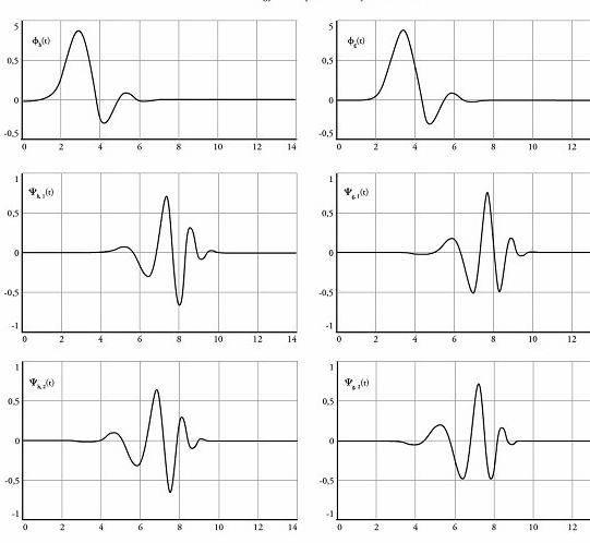
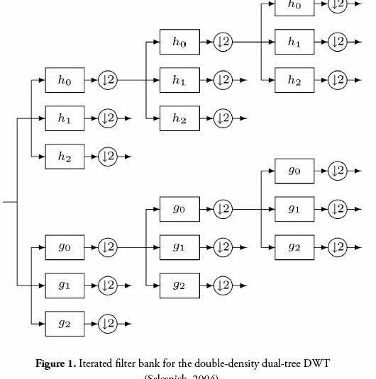
Within the second stage of lung lobe segmentation, stationary 2-D dual tree complex e wavelet transform [12] can be used identification in the particular fissure locations and curvatures inside the identified fissure regions helpful for that lobe segmentation formula. Isotropic CT images is challenging task for Identification of actual fissures regions because of the variable shape and appearance, coupled with low contrast and noise associated with these images. The thinness within the fissure is noted to be friends with 2 pixels (about 1.2 mm) in space, for the identification. In comparison, the two-D dual tree complex wavelet transform (DTCWT) boosts the contrast in the fissure region, while reducing the noise over the fissure. This produces a continuous line for the fissure. The multilevel decomposition within the DTCWT can be used picking out a an ideal amount of filtering (Fig 3.3). For this reason, the orthogonal wavelet filter can be used growing the perimeters within the fissures. Like the DWT, the DTCWT could be a multi resolution transform with decimated subbands offering perfect renovation within the input. In comparison, it uses analytic filters instead of real ones and so overcomes problems within the DWT at the expense of moderate redundancy. Running by 50 % DWT trees real and complex (real and imaginary) of real filters [11], this transform produces real and imaginary parts of the coefficients. The DTCWT decomposition plan’s proven in Fig 3.2.
In image processing technique, the stationary DWT uses low-pass and-pass filters to concurrently decompose the look, producing the approximation and detail coefficients in the image [7]. A Couple of-D DTCWT implies 1-D DTCWT to change the posts along with the rows. Unlike conventional wavelet transformations, the stationary 2-D DTCWT yields four sub images includes a low-pass kind of the very first image and three high-pass filtered images: horizontal, vertical, and diagonal. Further technique DTCWT for that low-pass image yields bigger levels of decompositions [8]. This space invariance property enables our formula to obtain the fissure location and curvature when using the detail coefficient images. To be able to comprehend the fissures, our formula applies a dual tree complex wavelet transform for that found fissure regions. One of the four images produced with this particular transform (low-pass, horizontal, vertical, and diagonal), the formula uses the horizontal detailed image for further processing since most lobar fissures appear horizontally inside the fissure regions. For the reason that identify the modified adaptive
g j = tan-1 æçç k j -k j-1 ö÷÷ è lj -lj-1 ø
fissure sweep that orients the fissure regions coupled with direction within the fissures. The formula uses second-level decompositions to create the horizontal detailed images because this level offers the best contrast for the fissures. To understand the particular fissure locations and curvatures, the lobe segmentation formula uses fissure search approach to uncover a extended continuous lines crossing the fissure regions, which signifies the particular fissures. The fissure search technique conducts pixel-by-pixel analysis and instantly placing anchor points a lengthy way away of 5 pixels apart along to understand the fissures. Transporting out a fissure search, the formula helpful for fissure verifies technique, which validates the correctness from the present fissure by evaluating its anchor points employing their counterparts round the previous adjacent fissure. Fig.3.2 describes the flow diagram for fissure search and fissure verification.
Since changes between fissures by 50 % adjacent isotropic CT images are extremely small, the next criteria define a highly effective fissure
Where g j (j = 1, 2, 3) may be the position within the fissure segment between two adjacent anchor points and possesses the type of
The lobe segmentation formula might be useful for your automated recognition within the fissure locations and curvatures for left oblique, right oblique and horizontal fissures are extracted.
IV RESULT AND DISCUSSION
The wavelet image de-noising using both DWT along with the DT-CWT for several wavelet functions remains put on selected CT lung pictures of database collected by us. The primary reason with this particular method is inside the recognition of specific biomedical image components in their beginning additionally for their visual enhancement. The DWT and DT-CWT both can be used lung images to extract the lobar fissure. Nevertheless the acquired result shows (in Fig 4) the later outplays the last in relation to precision within the extraction. The formula could properly comprehend the fissures under various pathological conditions. The little variation within the precision one of the patients was mainly because of the anatomic variations in the bronchi as well as other pathologies which have been present. Noise, pixel nonuniformity, poor contrast, and incomplete fissures mainly contributed incorrectness found in the fissures.
In Fig 4.a shows original CT Slice, Fig 4.b shows the thresholded image by equation(1), Fig 4.c shows the extracted lung region, Fig 4.d shows the elevated denoised lung, Fig 4.e shows the elevated bronchial trees and Fig 4.f shows the extracted left oblique fissure.
Fig.3.2. Flow diagram within the fissure search and verify techniques.
Fig.4. (a) Original slice, (b) Threshold image, (c) Extracted lung,
Enhanced denoised lung, (e) Enhanced bronchial trees,
Segmented left oblique fissure
Excellent, segmentation and analysis of CT (Computerized Tomography) images are very important tasks in diagnosing several illnesses. Variety and amount of images and sub images, throughout a particular situation, require involve efficient automatic methods for properly identify and segment abnormalities to make certain that further greater level processing might be transported out. Using dual-tree complex wavelet transform (DT-CWT), you are able to considerably reduce the amount of incorrectly detected points and to improve segmentation stage due to the shift-invariance and good directionality of DT-CWT.
Request Removal
If you’re the very first author in the essay with no longer want the essay printed across the United kingdom Essays website then please go here below to request removal:
More from United kingdom Essays




 Sample computer engineering thesis proposal
 Approval letter for thesis proposal
 Chemically modified electrodes thesis proposal
 Someones mother by joan murray thesis proposal
 Freire banking concept of education thesis proposal
